HM Hospitales ha dado un nuevo paso en la vanguardia tecnológica al servicio del paciente al realizar en un centro del grupo la primera intervención de cáncer de próstata en el grupo mediante la utilización de ultrasonidos de alta intensidad focalizados (HIFU), un tratamiento no invasivo de eficacia confirmada y numerosas ventajas para el paciente.
La tecnología HIFU trata el cáncer de próstata concentrando ultrasonidos de alta intensidad que destruyen las células de la glándula prostática mediante la aplicación de calor, sin afectar a los tejidos circundantes, explican los doctores Ramón Diz y Francisco Begara, urólogos del Hospital Universitario Madrid (HM).
“El mecanismo de acción de los ultrasonidos de alta intensidad es similar al de los rayos del sol cuando pasan a través de una lupa: todos los rayos luminosos se concentran en un punto y provocan un importante incremento de la temperatura alrededor del punto focal”, señalan, indicando que el HIFU permite la destrucción coagulativa del tejido prostático sin necesidad de una intervención quirúrgica abierta.
-Ventajas y eficacia de la técnica
Las ventajas del HIFU para el paciente, en opinión de estos expertos -que han intervenido al primer paciente de HM Hospitales tratado con esta técnica-, son claras: “Se trata de un tratamiento no invasivo que posibilita la rápida reincorporación del paciente a su vida cotidiana, permite la repetición del mismo y es personalizado”. Éste puede ser radical o focal, continúan: “Es posible decidir realizar una tratamiento radical de toda la glándula o emplearlo sólo en la zona afecta de ésta, con la intención de preservar la máximo la calidad de vida del paciente y limitando el impacto de la energía aplicada sobre los tejidos circundantes”. El hecho de ser un tratamiento robotizado, lo que implica precisión y seguridad, completa el listado de beneficios de esta técnica.
En cuanto a su eficacia, se mide en términos del nadir del PSA -valor más bajo del antígeno prostático específico (PSA) alcanzado tras cualquier tratamiento para el cáncer de próstata-, que se ha comprobado que después de la aplicación de la tecnología HIFU se correlaciona de forma altamente significativa con la supervivencia libre de enfermedad, aseguran ambos especialistas, que añaden que los resultados de la técnica mejoran si se alcanza un nadir menor o igual a 0,2 ng/ml siendo la supervivencia teórica a los cinco años del 86 por ciento en pacientes con un nadir de PSA menor de 0,5 ng/ml.
Por su parte, según los datos aportados por los doctores Diz y Begara, con la aplicación de HIFU se alcanza un índice de biopsias negativas del 92 por ciento en pacientes con gleason -indicador que valora el grado de actividad (agresividad y crecimiento) del cáncer de próstata- menor de 7, del 86 por ciento cuando el valor es de 7 y del 82 por ciento si el gleason es superior a ese nivel.
Adicionalmente, el tratamiento con esta técnica elimina problemas de incontinencia en el 98 por ciento de los pacientes, mientras que el 87 por ciento conservó su función sexual cuando se empleo el HIFU con una estrategia de preservación de los nervios erectores. “En los pacientes tratados tras radioterapia se obtiene un índice de biopsias negativas del 73 por ciento”, apostillan los urólogos.
-Indicaciones de HIFU
Esta técnica, cuya utilización se indicó inicialmente en pacientes con cáncer de próstata no candidatos a cirugía por co-morbilidad asociada o por negativa del paciente a someterse a cirugía radical prostática, ha ampliado su uso en la actualidad, estando indicada en pacientes que todavía no han sido tratados y bien tengan tumor localizado y en estadio T1 o T2, tengan un índice gleason inferior o igual a 7, o deseen conservar una calidad de vida optima tras el tratamiento.
Asimismo, los pacientes que busquen un enfoque terapéutico innovador con un tratamiento focal de la enfermedad que persiga un control de la misma a través de un estricto seguimiento, y con la posibilidad de repetir el procedimiento; aquellos que hayan experimentado recidiva local del cáncer tras su tratamiento con radioterapia externa, braquiterapia o bloqueo hormonal; los casos de tumores localmente avanzados -en estos cuadros la técnica HIFU se aplica como terapia adyuvante de reducción de masa tumoral local-; y los pacientes con recidiva local tras la prostatectomía radical son otros perfiles indicados para el uso de ultrasonidos de alta intensidad focalizados.
Por el contrario, los expertos señalan también situaciones en las que el empleo de esta técnica está contraindicado, tales como en las cirugías del recto o del ano que impidan la introducción de la sonda rectal, cuando el paciente tiene un esfínter artificial o una próstesis de pene, o ante la presencia de daño en la pared rectal por tratamientos previos rectales o de la próstata.
-Resultados y evolución muy positivos
El paciente intervenido el pasado mes de julio es un varón de 61 años diagnosticado en 2004 de cáncer de próstata gleason 3+3, que fue tratado inicialmente con braquiterapia y posteriormente, en 2011, causó recidiva bioquímica confirmada con una biopsia. “Al paciente se le ofrecieron todas las opciones terapéuticas viables para su situación y disponibles en HM Hospitales: cirugía radical abierta, laparoscópica y robótica, crioterapia e HIFU” eligiéndose finalmente esta última”, detallan los urólogos.
“El resultado de la intervención y la evolución del paciente han sido muy positivos -continúan-: se le dio de alta al día siguiente de la operación, sus niveles de PSA han vuelto a la normalidad y en la actualidad únicamente sigue revisiones con controles periódicos”.
Con la aplicación de ultrasonidos de alta intensidad focalizados la destrucción del tejido prostático se produce a través de tres mecanismos físicos diferentes: un efecto mecánico, un efecto térmico y la cavitación. Así, para los doctores Diz y Begara, los parámetros más importantes en la aplicación del HIFU son “la frecuencia de los ultrasonidos, la intensidad acústica, la duración de la aplicación, los intervalos de los pulsos, la distancia lateral entre las lesiones elementales, el desplazamiento longitudinal de la fuente de energía cuando se aplican múltiples lesiones y la profundidad de penetración, que dependerá del diseño del aplicador”.
-Precisión milimétrica
La técnica, en la que puede emplearse anestesia raquídea o general y requiere del paciente en posición decúbito lateral, utiliza una sonda que permite al especialista visualizar la glándula prostática, al tiempo que se emiten los ultrasonidos que destruirán el tejido prostático. “Gracias al programa informático que se usa el urólogo es capaz de visualizar los límites de la próstata, definir los márgenes de seguridad y planificar cada etapa del tratamiento con una precisión milimétrica”, explican los expertos.
Al término de la intervención, que puede durar entre una hora y media y dos horas y media, y en la que se realizan entre 400 y 800 pequeñas lesiones contiguas para tratar toda la próstata, se emplaza una sonda vesical que el paciente debe portar durante dos semanas.
Según indican los urólogos, las lesiones inducidas por el HIFU se identifican temporalmente como áreas hiperdensas en la ecografía transrectal de la glándula prostática. “La extensión del daño tisular provocado por el HIFU puede también determinarse con resonancia magnética con gadolinio: el área tratada aparece como una zona hipodensa rodeada por un borde periférico fuerte de 3-8 mm”, añaden, señalando, no obstante, que “los daños de RNM inducidos por el tratamiento desaparecen normalmente en 3-5 meses y la contracción del tejido inducido por el HIFU es el resultado obtenido al cabo aproximadamente de medio año”.
13 September 2011
Key signal that prompts production of insulin-producing beta cells points way toward diabetes cure

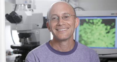
Researchers at the Hebrew University of Jerusalem have identified the key signal that prompts production of insulin-producing beta cells in the pancreas -- a breakthrough discovery that may ultimately help researchers find ways to restore or increase beta cell function in people with type 1 diabetes. The work on the multi-year project was led by Prof. Yuval Dor of the Institute for Medical Research Israel-Canada of the Hebrew University, researchers from the Hadassah University Medical Center and researchers from the diabetes section of the Roche pharmaceuticals company. The study was published in a recent issue of the journal Cell Metabolism.
"Our work shows that as the glucose level is increased in the blood, it tells the beta cells to regenerate," says Dor. "It's not blood glucose per se that is the signal, but the glucose-sensing capacity of the beta cell that's the key for regeneration." This was the first time that this sensing of a high level of glucose has been shown to be the "trigger" that induces beta cells to regenerate.
In persons suffering from type 1 (juvenile) diabetes, the immune system launches a misguided attack on the insulin-producing beta cells, resulting in the cells' decline of insulin production and eventual loss of function.
Without insulin, the body's cells cannot absorb glucose from the blood and use it for energy. As a result, glucose accumulates in the blood, leaving the body's cells and tissues starved for energy. That's why people with the disease must inject insulin and monitor their blood glucose levels diligently every day. To cure type 1 diabetes, it will be necessary to develop methods to increase beta cell replication and mass, hence the potential therapeutic importance of the current study.
In their work, Dor, along with co-lead author Prof. Benjamin Glaser of the Hadassah University Medical Center, used a genetic system to destroy 80 percent of the insulin-producing cells in the pancreases of adult mice, rendering the mice diabetic.
When the researchers compared these mice with control mice, they found that those mice with diabetes and elevated blood glucose levels had regenerated a greater number of new beta cells than mice without diabetes, suggesting that glucose may be a key player in beta cell regeneration. But the researchers further found that a glucose-sensing enzyme in the cells, glucokinase, is the key molecule that triggers the beta cell regeneration.
"This means that the more work that beta cells are required to do (that is, the more 'stressed' they are), the more of themselves they make," said graduate student Shay Porat, who, along with fellow graduate student Noa Weinberg, spearheaded the study, which was funded with the support of the Juvenile Diabetes Research Foundation (JDRF). .
Because this study showed that regeneration depends on glucokinase levels, the finding may pave the way for developing a new kind of drug to modulate glucokinase or other steps in the glucose-sensing pathway to direct beta cells down the path of regeneration and replication.
And, should a mechanism be discovered that prevents the immune system from attacking beta cells in the first place, as occurs among diabetics, the combined treatment could help pave the way towards a full cure for type 1 diabetes.
Further research in this area is proceeding, with the eventual goal of progressing towards human clinical trials.
*Source: The Hebrew University of Jerusalem
Reportaje en EL PAIS: Operaciones de obesidad sin bisturí

Son la última expresión de las cirugías mínimamente invasivas, en este caso aplicadas a la reducción de estómago: las operaciones sin bisturí ni hilo de sutura, dos de los ingredientes que parecen indispensables en cualquier quirófano. Y con resultados palpables y, sobre todo, pesables: los 19 kilogramos que ha perdido Enrique Lucini en el mes que ha pasado desde que se sometió a una intervención de este tipo.
"No lo hice por coquetería; fue una medida preventiva", recuerda Lucini. El hombre, madrileño de 49 años -aunque vive en Tenerife-, mide 1,85 metros y pesaba 131 kilogramos. "Estaba relativamente bien, la analítica era buena. Lo único que tenía eran apneas y unos ronquidos salvajes. Pero uno se acerca a los 50 años y tiene que tomar precauciones", dice.
De todas las opciones que se le plantearon para perder peso, eligió la cirugía de obesidad primaria por endoscopia. En inglés queda mucho mejor: las siglas forman la palabra POSE, la última novedad en este tipo de operaciones, como explica el médico Adelardo Caballero. "Es tan nueva que lleva menos de un año haciéndose en el mundo. En España solo la ofrecen la clínica Teknon de Barcelona y nosotros", dice el médico quien, como otros facultativos que trabajan en la sanidad privada, reparte su tiempo entre la clínica USP San José, la de La Luz (en Madrid) y una propia, que es donde, en este caso, hace el seguimiento del paciente.
"Nosotros todavía operamos con los americanos que inventaron la técnica aquí. De alguna manera estamos todavía en fase de entrenamiento. Nuestro objetivo es ofrecer la máxima eficacia con el menor riesgo", apunta el especialista. En los tres meses que hace desde que empezaron a operar con esta técnica ya han intervenido a una veintena de personas.
La operación parece sencilla. Una vez anestesiado el paciente, se le introduce por la boca un endoscopio que tiene en su extremo una especie de pinza que sirve para poner grapas en el estómago. "Se pinza hacia dentro, de manera que lo que se deja en contacto es la parte de fuera del estómago, que cicatriza y se une; así no hay peligro, como en otras intervenciones, de que se vuelva abrir el estómago, porque lo que se pone en contacto no es la mucosa, que pega muy mal", indica el médico mientras dobla un papel para explicar de manera gráfica el proceso que se aplica a esta cirugía.
Así contado parece fácil. "Pero hay que saber dónde y cuánto grapar", matiza el médico. En el caso de Lucini necesitó 15 grapas, y la intervención duró apenas 45 minutos. "En algunas llegamos a la hora y media, pero no es lo normal. Según vayamos perfeccionando la técnica el tiempo irá bajando", dice Caballero.
Con este modelo de operaciones se consigue disminuir la capacidad del estómago del paciente, que es la base de todas las intervenciones de reducción de este tipo. Pero se hace de una manera muy selectiva. "Cerramos sobre todo el lumen [la parte superior del estómago, la más cercana a la entrada del esófago]", explica el médico. Y, al intervenir ahí, se obtiene un efecto añadido, porque es en esa zona donde se produce la grelina, que es la hormona que cuando llega al cerebro le da la señal de que el paciente tiene apetito. "Al reducirla, se produce un efecto saciante", explica el cirujano.
El paciente confirma este efecto: "Vengo de pedirme un arroz con gambas y setas que no se lo saltaba un gitano, pero me he dejado la mitad y no me ha costado nada. Antes, eso hubiera sido imposible", cuenta Lucini.
Además, la técnica tiene la ventaja de que el posoperatorio es muy corto. "Estuve ingresado una noche, y eso porque me habían puesto anestesia general", dice Lucini. "Al día siguiente, salió del hospital y cogió un avión para Tenerife", remacha orgulloso Caballero. "Solo tuve algunos espasmos al volver a comer", relata el paciente.
Casi la mayor pega es el precio: unos 12.000 euros le ha costado todo el proceso a Lucini, ya que esta intervención no la ofrece la sanidad pública. "Ahora estoy de papeleos a ver si el seguro me lo paga", dice. Las molestias son tan escasas que en algunos casos el médico tiene que recurrir a trucos para que el paciente sea consciente de que le han hecho algo y que tiene que tomar precauciones. A las cinco semanas el paciente vuelve a hacer dieta normal.
Pero la relación con el médico no acaba con el alta. "La intervención no es un hecho aislado. Al paciente se le pone inmediatamente un programa de seguimiento de dos años, con psicóloga, nutricionista y entrenador personal que le va diciendo qué ejercicios debe hacer. Si tiene alguna duda, puede consultarnos por correo electrónico", dice Caballero.
Aprovechando la visita del paciente a Madrid, el médico le hace una revisión más completa. Aparte de pesarle y medirle la grasa corporal y el agua, con una ecografía le observa el hígado. "Lo tenía graso, que es síntoma de daño hepático, pero está reduciéndose", dice satisfecho.
El programa de seguimiento es clave. Tanto, que el médico afirma que si durante la entrevista que tiene con alguien interesado no le ve dispuesto a seguirlo, no le opera. "Rechazo hasta un 30% de las solicitudes", afirma Caballero.
Lucini no lo entiende. "Seguirlo es muy fácil. Claro que yo siempre he hecho mucho ejercicio. Juego al pádel a diario, al tenis, buceo, camino a diario. Pero me había descuidado. La diferencia es que ahora me canso menos, y disfruto mucho más. Ahora me miro en el espejo y no me reconozco. Y eso que aún me quedan 17 kilos por perder", dice seguro de que lo conseguirá.
**Publicado en "EL PAIS"
Even low-dose aspirin may increase risk of GI bleeding
The risk of gastrointestinal (GI) bleeding needs to be considered when determining the potential preventive benefits associated with low-dose aspirin for cardiovascular disease and cancer. According to a new study in Clinical Gastroenterology and Hepatology, the use of low-dose aspirin increases the risk for GI bleeding, with the risk being increased further with accompanying use of cardiovascular disease-preventing therapies, such as clopidogrel and anticoagulants. In patients who took proton pump inhibitors (PPIs), bleeding risk decreased. "The use of aspirin has been proven beneficial in reducing cardiac events and deaths in patients who have cardiovascular disease, and has even been shown to reduce cancer risk," said Angel Lanas, MD, PhD, of University Hospital Lozano Blesa and lead author of this study. "However, clinicians need to be more proactive in their efforts to reduce potential risk factors associated with all doses of aspirin, especially gastrointestinal bleeding. New low-dose aspirin studies should report more precisely on the incidence of bleedings, especially gastrointestinal bleedings, to better determine the balance between risks and benefits ."
Low-dose aspirin -- commonly defined as 75 to 325 mg daily -- is a mainstay of therapy for cardiovascular disease. In fact, patients with prior cardiovascular disease have fewer cardiovascular events and deaths with the use of low-dose aspirin compared with patients who do not use it. It is now likely to also be used for cancer prevention, especially GI and colon cancer.
A major factor limiting the widespread use of aspirin is concern about the development of GI adverse events, especially GI bleeding. However, damage may vary depending on the dose taken, other medication being consumed along with aspirin and patients' risk profiles. For example, certain patients have an increased likelihood of experiencing bleeding: those with long-term pharmacotherapy use, patients using combinations of low-dose aspirin with clopidogrel and anticoagulants, and patients with previous GI ulcers or bleedings.
In this study, doctors searched 10 electronic databases and collected data on adverse events in studies that evaluated low doses of aspirin alone or in combination with anticoagulants, clopidogrel or PPIs. They found that low doses of aspirin alone decreased the risk of death. However, the risk of major GI bleeding increased with low doses of aspirin alone compared with placebo. The risk also increased when aspirin was combined with clopidogrel (compared with aspirin alone), anticoagulants versus low doses of aspirin alone, or in studies that included patients with a history of GI bleeding or of longer duration. Importantly, PPI use reduced the risk for major GI bleeding in patients given low doses of aspirin.
**Source: American Gastroenterological Association
Fish oil reduces effectiveness of chemotherapy
Researchers at University Medical Center Utrecht, the Netherlands, have discovered a substance that has an adverse effect on nearly all types of chemotherapy -- making cancer cells insensitive to the treatment. Chemotherapy often loses effectiveness over time. It is often unclear how or why this happens. It now appears that chemotherapy is made ineffective by two types of fatty acid that are made by stem cells in the blood. Under the influence of cisplatin chemotherapy, the stem cells secrete these fatty acids that induce resistance to a broad spectrum of chemotherapies. These substances are referred to by researchers as 'PIFAs' which stands for platinum-induced fatty acids. Cisplatin is a type of chemotherapy that is widely used for the treatment of cancer, including cancer of the lungs and ovaries.
Tumors under the skin
The researchers studied the effect of PIFA's in mice and human cells. The mice studied had tumors under the skin. Under normal conditions, the tumors would decrease in size following the administration of chemotherapy. In the study, after administering the fatty acids to the mice, the tumors were found to be insensitive to chemotherapy. The fatty acids were isolated from the medium in which chemotherapy exposed stem cells were grown. But also stem cells in the blood of patients produce the fatty acids that desensitize tumors to chemotherapy.
The fatty acids are also found in commercially-produced fish oil supplements containing omega-3 and omega-6 fatty acids as well as in some algae extracts. In the experiments conducted in mice, the tumors became insensitive to chemotherapy after administration of normal amounts of fish oil. Natural products that include fish oil are frequently used by cancer patients in addition to their regular treatment.
"Don't use these products"
Professor Emile Voest, a medical oncologist at UMC Utrecht, supervised the research. "Where resistance to chemotherapy is concerned, we usually believe that changes in the cancer cells themselves have occurred. Now we show that the body itself secretes protective substances into the blood that are powerful enough to block the effect of chemotherapy. These substances can be found in some types of fish oil. Whilst waiting for the results of further research, we currently recommend that these products should not be used whilst people are undergoing chemotherapy."
Researchers at the University Medical Center Utrecht, the Netherlands, describe these findings, that will appear online on September 12, in the journal Cancer Cell.
*Source: University Medical Center Utrecht
First proof in patients of an improved 'magic bullet' for cancer detection and radio-therapy
Oncologists have long sought a powerful "magic bullet" that can find tumors wherever they hide in the body so that they can be imaged and then destroyed. Until recently scientists accepted the notion that such an agent, an agonist, needed to enter and accumulate in the cancerous cells to act. An international research team has now shown in cancer patients that an investigational agent that sticks onto the surface of tumor cells without triggering internalization, an antagonist, may be safer and even more effective than agonists. One of the Salk Institute's leading researchers, Dr. Jean Rivier, professor in The Clayton Foundation Laboratories for Peptide Biology and holder of the Frederik Paulsen Chair in Neurosciences and his Swiss collaborator, Dr. Jean Claude Reubi, University of Berne and Adjunct Professor at Salk, co-authored a pilot study, published in the September issue of the Journal of Nuclear Medicine, of five patients and demonstrated that their "antagonist," 111In-DOTA-BASS, outperformed the "agonist" agent, OctreoScan, that is widely used in the clinic to image neuroendocrine tumors bearing somatostatin receptors.
"This is the first proof of principle in humans that labeled peptide antagonists can effectively image tumors. Additional research suggests that we could one day use a different radioactive metal to effectively kill the tumors," said Dr. Rivier.
Dr. Reubi, a molecular pathologist, and Dr. Rivier, a chemist, collaborated in the design and selection of natIn-DOTA-BASS for human testing, and Dr. Helmut R. Maecke, a radio chemist, loaded DOTA-BASS with its radioactive marker and tested the compound before use in human. Afterward, the "first in man" study with the radioactive loaded DOTA-BASS was performed at the University Hospital in Freiburgby Drs. Damian Wild, Melpomeni Fani, Martin Behe, Ingo Brink, Helmut R. Maecke, and Wolfgang A. Weber.
The genesis of this study goes back to 1973, when a team of Salk researchers, which included Drs. Brazeau, Vale, Burgus, Rivier, and Roger Guillemin, a 1977 Nobel laureate, isolated and characterized somatostatin, a peptide produced by neuroendocrine glands. The scientists found that the normal function of somatostatin is to block the release of growth hormone throughout the body, which includes inhibiting the release of thyroid-stimulating hormone (TSH) from the thyroid.
Drs. Rivier, Reubi and their colleagues from Germany showed that 111In-DOTA-BASS bound to a greater number of somatostatin receptors on cancer cells than the agonist OctreoScan, and that it did accumulate in normal tissue (liver and kidney) to a lesser extent.
The prototype antagonist therapy has been revamped, and the version studied in the Journal of Nuclear Medicine publication, 111In-DOTA-BASS, detected 25 of 28 metastatic neuroendocrine tumors in the patients, whereas OctreoScan detected only 17.
In-DOTA-BASS has been licensed to a pharmaceutical company for clinical trial development, according to Rivier, who adds that other researchers are exploring an antagonist approach for other G-protein coupled receptors that are abundantly expressed on cancer cells.
The study was funded in part by the Swiss National Science Foundation (JCR).
**Source: Salk Institute
Ophthalmic antibiotics associated with antimicrobial resistance after intraocular injection therapy
Repeated exposure of the eye to ophthalmic antibiotics appears to be associated with the emergence of resistant strains of microbes among patients undergoing intraocular injection therapy for neovascular retinal disease, according to a report in the September issue of Archives of Ophthalmology, one of the JAMA/Archives journals. According to background information in the article, more than 8 million people in the United States are affected by age-related macular degeneration, the leading cause of blindness among individuals older than 65 years in this country. Treating the neovascular or "wet" form of the disease involves monthly injections of medication into the eye; this treatment is also being studied for eye problems related to diabetes and retina vein occlusions (obstructions of veins carrying blood from the retina). To prevent the most severe complication from intraocular injection, endophthalmitis (inflammation inside the eye), ophthalmologists routinely prescribe ophthalmic antibiotics after every injection. "Repeated exposure of ocular flora [microbes living on or inside the body], however, may select for resistant bacterial strains and cultivate 'superbugs' with multiple-drug resistance that may considerably affect the treatment of ocular infections," write the authors.
Stephen J. Kim, M.D., and Hassanain S. Toma, M.D., from the Vanderbilt University School of Medicine, Nashville, Tenn., conducted a randomized, controlled, longitudinal study of 48 eyes of 24 patients who, in one eye each, received intraocular injection. At baseline and after every injection, researchers obtained cultures of the conjunctiva (the membrane of the eye's surface and the inner eyelid) for both treated and untreated (control) eyes. Patients were randomized to one of four antibiotics and after each injection used only the antibiotic they were assigned. The researchers tested the bacterial samples for susceptibility to 16 antibiotics and analyzed the bacterial DNA. Injections were administered every four weeks for at least four consecutive months, and patients were followed for one year.
Repeated exposure to fluoroquinolone antibiotics was associated with coagulase-negative staphylococci (CNS) that demonstrated significantly increased rates of resistance to both older- and newer-generation fluoroquinolones. Repeated exposure to azithromycin was associated with CNS that demonstrated significantly increased resistance to macrolides and decreased resistance to both older- and newer-generation fluoroquinolones. Specimens of CNS from treated eyes demonstrated significant increases in multiple-drug resistance; for example, 81.8 percent of CNS specimens appeared resistant to at least three antibiotics, and 67.5 percent appeared resistant to at least five antibiotics.
The researchers suggest that their results demonstrate rapid development of resistance from CNS to certain antibiotics, and that this resistance is maintained when the antibiotic is periodically readministered. "This finding has considerable implications because conjunctival flora are presumed to be the predominant source of postinjection endophthalmitis," they write, adding that research suggests one strain of CNS is associated with greater intraocular inflammation than are strains more susceptible to antibiotics. "Our findings," the authors conclude, "indicate the need for more judicious use of ophthalmic antibiotics after intraocular injection to reduce the potential emergence and spread of antimicrobial resistance."
*Source: JAMA and Archives Journals
Subscribe to:
Posts (Atom)